What Is Cerebral Folate Deficiency
Folates are a family of more than 25 structurally related compounds collectively known as vitamin B9. They are essential for DNA synthesis, cell division, neurotransmitter production, and epigenetic control of gene expression. Without adequate folate, several critical processes in the nervous system break down at once: dopamine and serotonin synthesis, DNA methylation, myelin production, and antioxidant defense.
In 2004, a research group led by Vincent Ramaekers coined the term cerebral folate deficiency (CFD). It describes a condition in which the level of the active folate form — 5-methyltetrahydrofolate (MTHF) — is low in the cerebrospinal fluid, even though blood folate, vitamin B12, and homocysteine levels remain normal.
This is the key point: a standard blood test for folate and B12 will not detect cerebral deficiency. The brain can be starving while the blood test looks perfectly normal.
How Folate Reaches the Brain
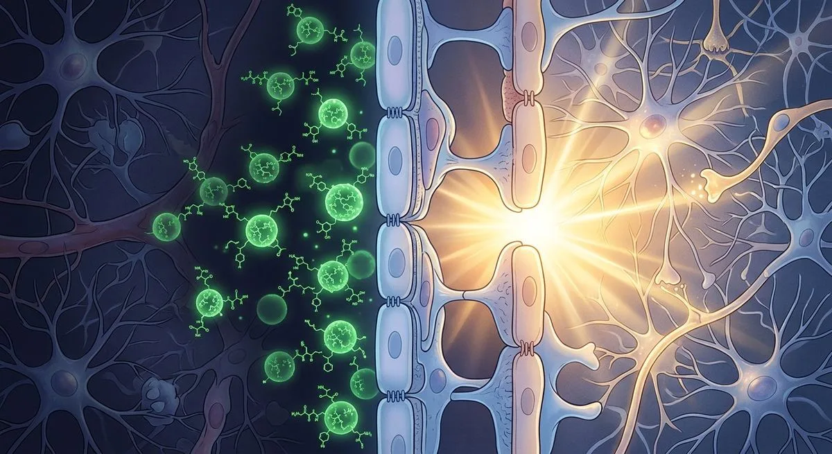
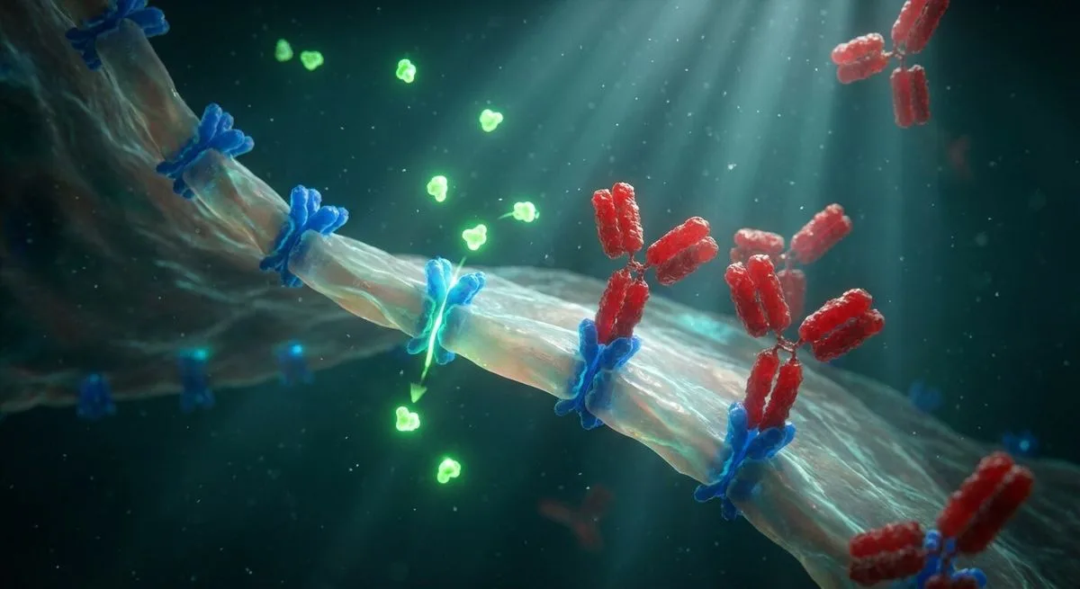
The main form of folate circulating in plasma is 5-methyltetrahydrofolate (MTHF). To reach the brain, MTHF must cross the blood-brain barrier at the choroid plexus. This transport is carried out by two proteins working together:
- Folate receptor alpha (FRα) — a membrane protein anchored to the surface of choroid plexus cells via a GPI anchor. It binds MTHF and triggers endocytosis — pulling the molecule into the cell inside a vesicle.
- Proton-coupled folate transporter (PCFT) — releases folate from the endosome into the cell.
After crossing the choroid plexus, MTHF enters the cerebrospinal fluid, then passes through the ependymal barrier into brain parenchyma and is taken up into neuronal metabolism.
The same FRα-mediated transport mechanism operates at the placental barrier (delivering folate to the fetus), in the gonads (both male and female), and in the thyroid gland during the prenatal period and the first months after birth.
Five Mechanisms of Cerebral Folate Deficiency
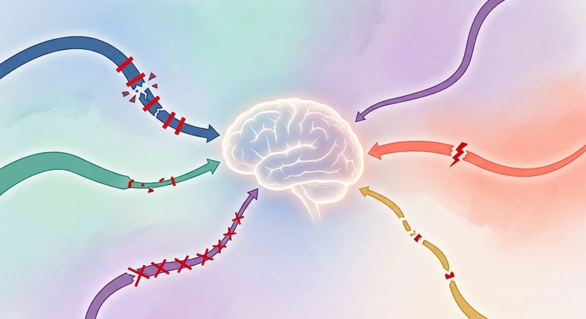
According to the classification proposed by Ramaekers and Quadros, five main mechanisms lead to CFD:
I. Impaired folate transport across the blood-brain barrier. This includes FRα dysfunction (autoantibodies, mutations in the FOLR1 gene, disrupted endocytosis signaling pathways, altered membrane composition in Smith-Lemli-Opitz syndrome), impaired energy metabolism (mitochondrial disease, GLUT1 deficiency), and damage to the choroid plexus (hemorrhage in premature infants, infectious and immune-mediated injury, oxidative stress).
II. Depletion of the intracellular folylpolyglutamate pool. A reduction in the stored forms of folate inside neurons.
III. Increased folate consumption in the brain. Inherited enzyme deficiencies (aromatic L-amino acid decarboxylase deficiency, dihydropteridine reductase deficiency), iatrogenic causes (carbidopa in Parkinson's disease treatment), and infectious or immune-mediated processes (subacute sclerosing panencephalitis, Rasmussen encephalitis).
IV. Increased folate catabolism. Oxidative and nitrosative stress break down MTHF molecules and damage transport proteins. Deficiency of antioxidant enzymes (glutathione peroxidase, superoxide dismutase) and their cofactors (selenium, manganese, coenzyme Q10) makes the situation worse.
V. Disorders of brain folate metabolism. Deficiencies of folate-cycle enzymes (MTHFR, serine hydroxymethyltransferase, dihydrofolate reductase), depletion of the one-carbon donor pool (3-phosphoglycerate dehydrogenase deficiency), and hyperhomocysteinemia caused by combined deficiency of vitamins B2, B6, B9, and B12.
FRα Autoantibodies: The Leading Cause
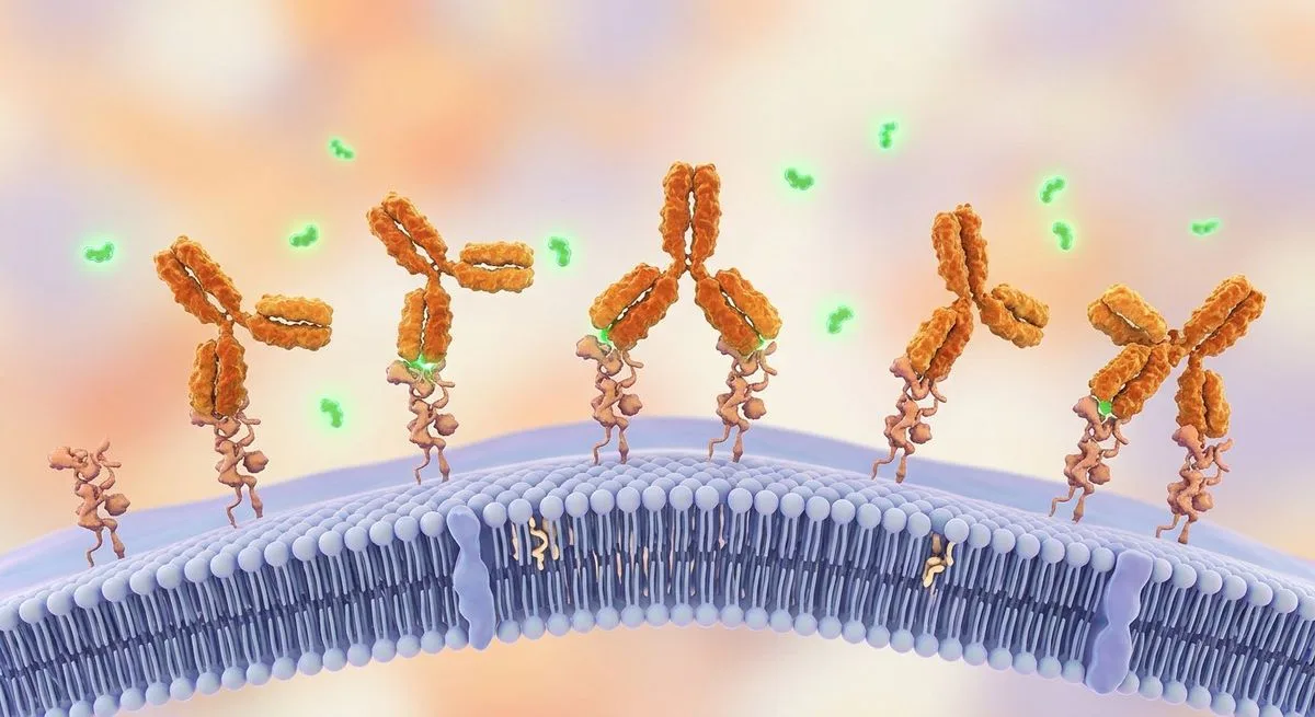
The most common cause of CFD is autoantibodies against folate receptor alpha (FRα). There are two types:
- Blocking antibodies — bind directly to the region of FRα responsible for capturing folate, physically preventing MTHF from binding.
- Binding antibodies — attach to FRα away from the folate-binding site but trigger complement-mediated inflammation that ultimately destroys the FRα-antibody complex.
Both mechanisms lead to the same outcome — reduced overall folate transport into the brain.
Where These Antibodies Come From
The human FRα protein shares roughly 90% amino acid sequence homology with the FRα found in cow's milk and other animal dairy products. In genetically predisposed individuals, soluble FRα absorbed from cow's milk through the gut stimulates the immune system to produce antibodies that cross-react with the body's own FRα in the choroid plexus, thyroid gland, and gonads.
This mechanism has experimental support: a dairy-free diet (eliminating milk and animal dairy products) lowers FRα antibody titers within 3-6 months, while reintroducing dairy causes titers to rise again.
An inverse correlation has been established between FRα antibody titer and MTHF levels in the cerebrospinal fluid. Another important feature: antibody titers fluctuate cyclically with a period of 5-7 weeks, so a single negative result does not rule out the diagnosis.
The Age Spectrum of Clinical Syndromes

The clinical picture of CFD depends on the age at which brain folate deficiency develops, as well as whether or not the parents have FRα autoimmunity.
Infantile CFD (Onset at 4-6 Months)
This is the first described and most thoroughly studied form. Initial symptoms — sleep disturbances, restlessness, and agitation in a 4-6-month-old infant — cannot be explained by gastroesophageal reflux or milk allergy. Over the following two years, the full clinical picture emerges:
- Slowed head circumference growth
- Psychomotor delay, hypotonia, ataxia
- Distal pyramidal signs (progressing to tetraspasticity if untreated)
- Dyskinesias (choreoathetosis, dystonia) and/or epileptic seizures in a third of patients
- Progressive vision loss starting at age 3, hearing loss starting at age 6
MRI in half of untreated patients shows delayed myelination, subcortical and periventricular demyelination, and cerebral and cerebellar atrophy. A minority of children with infantile CFD go on to develop autism.
Autism With Neurological Deficit
Children with low IQ, autism, and the typical neurological features of infantile CFD show cerebrospinal fluid MTHF levels and FRα antibody titers just as low as in "classic" infantile CFD. The key difference: one or both parents of children with autism test positive for FRα antibodies, whereas parents of children with infantile CFD but no autism do not.
This suggests that autistic features may stem from the consequences of parental FRα autoimmunity, which affects folate-dependent processes in sperm, oocytes, and/or fetal development.
Infantile Autism Without Neurological Deficit
Children with autism but no pronounced neurological impairment show a less severe drop in cerebrospinal fluid MTHF compared with infantile CFD, though levels are still statistically significantly lower than in healthy controls. FRα antibody titers are comparable to those in other groups. A substantial proportion of these children's parents also have FRα autoimmunity.
Spastic-Ataxic Syndrome (1-2 Years)
The emergence of FRα antibodies in children aged 1-2 years leads to a spastic-ataxic CFD syndrome accompanied by learning difficulties.
ADHD and Behavioral Disorders (2-5 Years)
A small proportion of children aged 2-5 with attention-deficit/hyperactivity disorder (ADHD), learning difficulties, and behavioral problems test positive for FRα autoimmunity. Neurological examination reveals mild gait ataxia and a positive Romberg test.
Schizophrenia and Psychotic Disorders (Adolescents and Adults)
FRα autoimmunity may predispose patients to severe psychotic episodes and treatment-refractory schizophrenia. In a study of 20 patients with refractory schizophrenia, FRα antibodies were found in 85% of cases. A characteristic feature is pronounced antibody titer fluctuation with a 5-7 week period, which may explain the alternation between positive and negative symptoms.
Treatment-Resistant Depression
FRα antibodies were found in 9 of 16 patients tested (56%) with severe, treatment-resistant major depression. Folinic acid therapy has shown some degree of effectiveness.
Dystonia and Parkinsonism
Sporadic cases have been reported of severe dystonia or parkinsonism combined with psychiatric disorders unresponsive to standard therapy, in which CFD driven by FRα autoimmunity was identified.
Dementia (Adults)
A case has been reported of an adult woman with dementia and myoclonus who was diagnosed with CFD syndrome. Systematic studies across different forms of dementia have not yet been conducted.
Diagnostic Algorithm
Diagnosing CFD starts with clinical suspicion, taking into account the patient's age, history, and neurological status.
First-Line Workup
- Complete blood count, serum and red-cell folate, plasma vitamin B12, homocysteine, lactate, CK
- Liver and kidney function, TSH, T3, T4
- Ferritin, copper, zinc, manganese, selenium
- Total cholesterol, apolipoprotein B, coenzyme Q10
- Vitamins A, B2, B6, C, D, E, and gamma-tocopherol
- Anti-gliadin antibodies
- MTHFR genotyping (C677T and A1298C)
The Key Test
Serum FRα autoantibody testing (both blocking and binding types). Important: folate-containing supplements should be stopped 3 days before testing. If the result is negative, retesting after a few weeks is recommended because of the cyclical nature of titer fluctuations.
For infantile CFD and autism, a lumbar puncture is not always required — serum FRα antibodies, which inversely correlate with cerebrospinal fluid MTHF levels, are checked first.
If FRα Antibodies Are Negative
- FOLR1 and CIC gene mutations
- Ruling out mitochondrial defects
- MTHFR mutations
- Oxidative stress markers and levels of antioxidant enzyme cofactors (iron, copper, zinc, selenium, manganese)
- Lumbar puncture to measure MTHF, pterins, and monoamine metabolites
For Autism — Testing the Parents
If a child with autism tests positive for FRα antibodies, testing both parents for antibodies is recommended — this affects the treatment prognosis.
Folinic Acid
The mainstay of treatment for CFD caused by FRα autoimmunity is high pharmacological doses of folinic acid:
- dl-folinic acid (dl-5-formyltetrahydrofolate): starting dose 0.5-1 mg/kg/day, increased to 2 mg/kg/day if needed (maximum 50 mg/day)
- Levo-folinic acid: 0.25-0.50 mg/kg/day
- Levo-5-methyltetrahydrofolate: an equivalent dose (not approved for medical use in Europe)
Critically important: treatment should start at half the target dose for the first month. A sudden rise in brain folate increases production of tetrahydrobiopterin, dopamine, and serotonin, leading to synaptic overstimulation. It takes about 6 weeks for a new balance to establish itself between neurotransmitter output and the density of previously suppressed receptors. For pronounced agitation or aggression, a low dose of risperidone may be prescribed temporarily for 2-3 months.
For CFD caused by FOLR1 mutations, higher doses — up to 5-7 mg/kg/day — are needed to compensate for the functional loss of the receptor.
Dairy-Free Diet
Eliminating milk and animal dairy products (replacing them with plant-based milk) removes exposure to the soluble FRα that triggers the cross-reactive immune response. FRα antibody titers drop after 3-6 months on the diet. Dietary changes can be introduced right after diagnostic samples are collected, without waiting for the results.
Limitation: children with autism spectrum disorders often refuse or cannot tolerate such a strict diet because of food selectivity.
Special Situations
Severe dyskinesias, frequent seizures, status epilepticus: corticosteroids to suppress FRα autoimmunity until folinic acid therapy takes effect.
Mitochondrial disease (Alpers disease, Kearns-Sayre syndrome): folinic acid produces a variable response; adding antioxidants may help, but convincing evidence of meaningful improvement is still lacking.
MTHFR mutations (C677T homozygous, C677T/A1298C compound heterozygous): it makes sense to replace part of the folinic acid dose with an equivalent dose of levo-methyl-THF, since MTHF is the only folate form that crosses the blood-brain barrier. High-dose riboflavin is needed to boost MTHFR activity.
Vitamin D deficiency: reduces expression of the RFC1 gene (an alternative folate transport pathway that operates at high concentrations). Correcting vitamin D deficiency is mandatory during CFD treatment.
Nutritional deficiencies in autism: systematic screening for deficiencies in vitamins C, E, gamma-tocopherol, coenzyme Q10, iron, copper, zinc, manganese, and selenium — all of these function as antioxidants or antioxidant enzyme cofactors, and their shortage worsens the oxidative stress that disrupts folate metabolism.
Looking Ahead: Prevention and Screening
Observational data show that children with infantile CFD who receive folinic acid treatment before age 2 have the best outcomes, up to full recovery. After age 6, the prognosis is substantially worse.
In autism with FRα autoimmunity, the prognosis depends on whether the parents also carry antibodies:
- Antibodies in the child only — the most favorable response to treatment
- Antibodies in the child and one parent — partial reversibility
- Antibodies in both parents — an unfavorable prognosis despite treatment
Preventing Autism in Siblings
Preliminary data from three families in which the first child had infantile autism with positive FRα antibodies:
The parents received low-dose folinic acid (up to 3.75 mg/day of levo-folinic acid), starting 3 months before conception. Mothers with positive FRα antibodies continued taking it throughout the pregnancy. Genetic testing (CNV analysis and known autism genes) in the first child was normal.
Five healthy siblings were born across the three families — preliminary data that still needs confirmation in clinical trials, but one that opens up the possibility of preventive screening and pre-emptive treatment in at-risk families.
Proposed Screening Measures
The review's authors propose considering:
- FRα antibody screening in mothers and in cord blood
- Monitoring the child for FRα antibodies at 6, 12, and 24 months
- Testing prospective parents for FRα antibodies and associated genetic variants before conception
These proposals require dedicated clinical studies to confirm the effectiveness of early intervention.