
The Look-Alike Protein
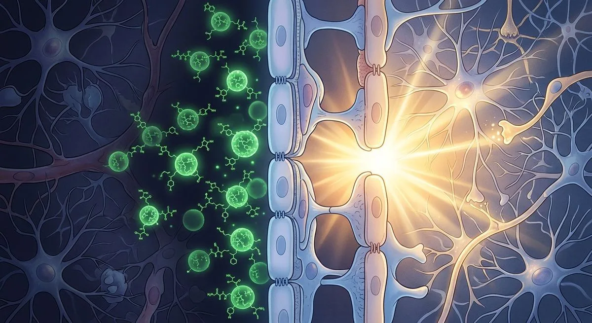
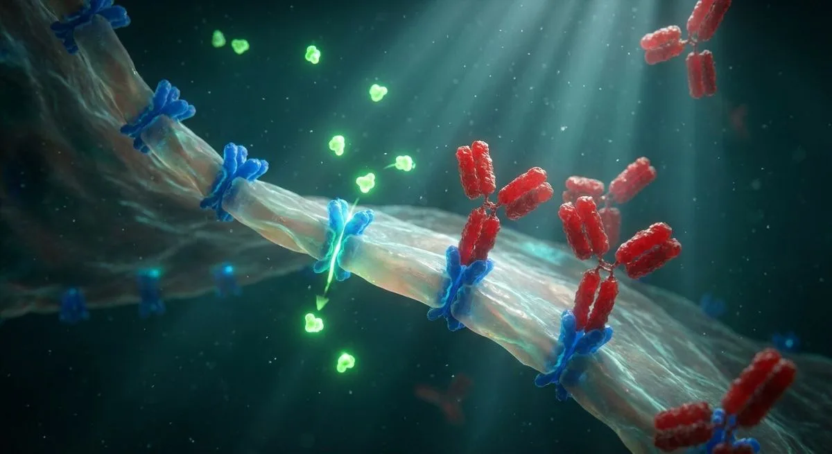
Cow's milk contains a protein that rarely gets talked about. It's called folate receptor alpha — FRα. The very same protein sits on the surface of choroid plexus cells in the human brain. Its job is to capture vitamin B9 (folate) from the blood and ferry it across the blood-brain barrier into the nervous system.
Human and bovine FRα are roughly 90% identical in amino acid sequence. To the immune system, they're practically the same protein.
For most people, that never causes a problem. But in those who are genetically predisposed, it sets off a chain reaction: the immune system recognizes the FRα from cow's milk as a foreign protein, produces antibodies against it, and those antibodies then turn on the body's own FRα at the blood-brain barrier. The gate that lets folate into the brain swings shut.
How It Works
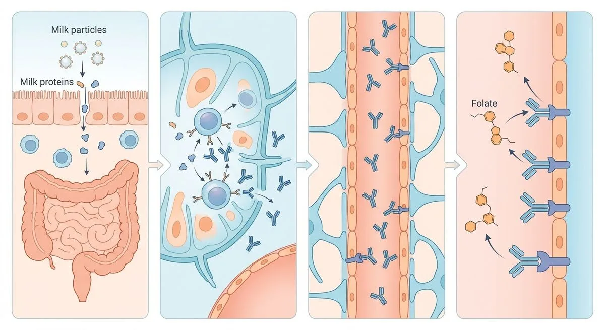
The mechanism unfolds in four steps.
Step 1: Exposure. A child or adult consumes cow's milk and dairy products. Soluble FRα from the milk enters the gut and then the bloodstream.
Step 2: Immune response. The gut immune system of a genetically predisposed person recognizes dairy FRα as an antigen and starts producing two types of antibodies. Blocking antibodies attach directly to the site on FRα that binds folate, physically preventing the vitamin B9 molecule from docking. Binding antibodies attach to FRα elsewhere, but they activate the complement system, which destroys the entire receptor-antibody complex.
Step 3: Cross-reactivity. Antibodies raised against dairy FRα cross-react with the body's own FRα on the choroid plexus, the thyroid gland, and the gonads — precisely because of that 90% homology.
Step 4: Deficiency. Folate transport across the blood-brain barrier drops. The brain is starved of 5-methyltetrahydrofolate (MTHF) — the active form of vitamin B9 — even though blood folate levels stay normal. A standard blood test misses the problem entirely.
There's direct evidence for this mechanism: removing milk and dairy products of animal origin from the diet causes FRα-antibody titers to drop within 3-6 months. Reintroduce milk, and the antibodies climb again.
A Starving Brain — What Happens
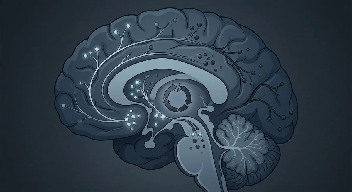
Folate in the brain isn't just "a vitamin." It's a molecule that several critical biochemical pathways depend on simultaneously.
Neurotransmitters. MTHF takes part in a reaction chain that, through tetrahydrobiopterin (BH4), drives the synthesis of dopamine, serotonin, and nitric oxide. Without enough folate, production of the neurotransmitters that regulate mood, attention, motivation, and motor function falls.
Methylation and epigenetics. MTHF is a key methyl donor for synthesizing S-adenosylmethionine (SAM), the body's universal methylating agent. SAM drives more than 100 methylation reactions, including DNA and histone methylation. Folate deficiency leads to DNA hypomethylation, disrupted gene silencing, and impaired neuron maturation, neurite outgrowth, and neural network formation.
DNA synthesis. Folate is needed to synthesize thymidine and purines — the building blocks of DNA. Its deficiency disrupts cell division and DNA repair.
Antioxidant defense. Through the transsulfuration pathway, homocysteine is converted into glutathione — the main intracellular antioxidant. When folate is deficient, homocysteine builds up and glutathione levels fall. Oxidative stress rises, further damaging the remaining folate and its transport proteins. The vicious circle closes.
Not Just Autism

Cerebral folate deficiency (CFD) isn't a single disease but a spectrum of conditions. Which disorder actually develops depends on the age at which the antibodies start acting and on whether the parents themselves have FRα autoimmunity.
From birth — when the mother has antibodies. If the mother carries FRα antibodies, they disrupt folate transport across the placenta to the fetus. This raises the risk of neural tube defects, autism, and neurodevelopmental delay. Studies confirm that folate deficiency in early pregnancy correlates with autism in offspring.
4-6 months: infantile CFD. Sleep disturbances and irritability, followed by slowed head growth, developmental delay, hypotonia, ataxia, and pyramidal signs. About a third of affected children develop dyskinesias and seizures. Left untreated, vision loss can begin around age 3 and hearing loss around age 6.
1-2 years: spastic-ataxic syndrome. Movement and learning impairments.
2-5 years: ADHD. FRα antibodies are found in some children with ADHD, learning difficulties, and behavioral problems.
Adolescents and adults: psychiatric disorders. The data here are especially striking:
- Schizophrenia: FRα antibodies were found in 85% of patients with treatment-refractory schizophrenia (20 patients studied). The 5-7 week cyclical pattern of the antibodies may explain the alternating phases of relapse and remission.
- Depression: FRα antibodies were found in 56% of patients with severe treatment-resistant depression.
- Dystonia and parkinsonism: Sporadic cases that don't respond to standard therapy.
Older age: dementia. One case has been described of an adult woman with dementia and myoclonus linked to CFD. There are no systematic studies in dementia yet — but the case alone is worth pausing on.
The Blood Test No One Orders

Serum FRα autoantibody testing is a test that can explain the unexplainable. But it isn't part of any standard screening panel — it's only ordered when someone specifically suspects the condition.
There are a few nuances worth knowing:
Cyclicality. FRα antibody titers fluctuate on a 5-7 week cycle. That means a single negative result doesn't rule out the diagnosis — antibodies can be below the detection threshold today and spike sharply three weeks later.
Folate supplements. Folate-containing vitamin supplements need to be stopped 3 days before testing, or the result can be skewed.
Inverse correlation. The higher the FRα antibody titer, the lower the MTHF level in cerebrospinal fluid. This relationship is backed by data: at zero antibody levels, CSF MTHF reaches 60-95 nmol/L; at a titer of 4.0 pmol/mL, it drops to 5 nmol/L.
A lumbar puncture isn't always necessary. For infantile CFD and autism, testing can start with blood FRα antibodies — their presence and inverse correlation with brain folate levels are well established. This considerably simplifies diagnosis, especially in young children.
If FRα antibodies come back negative on repeat testing but clinical suspicion remains, other causes need to be considered: FOLR1 gene mutations, mitochondrial defects, MTHFR mutations, oxidative stress.
Folinic Acid

The backbone of treatment is high pharmacological doses of folinic acid (5-formyltetrahydrofolate). It bypasses the blocked FRα transport route, partly using the alternative RFC1 carrier.
Dosing: dl-folinic acid at 0.5-1 mg/kg/day, with a possible increase to 2 mg/kg/day (maximum 50 mg/day). Levo-forms are used at half those doses.
One important rule: start at half the dose and titrate up gradually over the first month. A sharp rise in brain folate triggers a surge in dopamine and serotonin production, and the receptors for them haven't yet adapted after a long period of suppression. The result can be pronounced agitation and aggression. Stabilization takes about 6 weeks.
A Dairy-Free Diet
Eliminating milk and dairy products of animal origin removes the source of the cross-reacting FRα. Antibodies decline within 3-6 months. This can be started right after diagnostic samples are collected.
Correcting Co-Occurring Deficiencies
Many children with autism are found to be deficient in vitamins and trace elements: C, E, coenzyme Q10, iron, copper, zinc, manganese, selenium. All of these play a role in antioxidant defense. Their deficiency worsens the oxidative stress that further damages folate and its transport proteins — a closed loop that has to be broken on multiple fronts at once.
Vitamin D deserves a special mention: its deficiency lowers expression of the RFC1 gene, the alternative folate transporter. Correcting vitamin D levels is a mandatory part of CFD therapy.
In patients with MTHFR mutations (C677T homozygous), part of the folinic acid dose is replaced with levofolinic acid (L-methylfolate), and high doses of riboflavin (B2) boost the enzyme's residual activity.
Three Families Who Changed the Statistics

This is preliminary data — but it deserves attention.
In three families, the first child had autism with positive FRα antibodies. Genetic testing for known causes of autism (CNVs, associated genes) came back normal. FRα antibodies were found in the mothers in all three families, and in the fathers in one of the three.
The parents began taking low-dose folinic acid (up to 3.75 mg/day of levofolinic acid) three months before conception. Mothers with antibodies continued taking it throughout pregnancy.
The result: five subsequent children across the three families — all healthy. Not a single case of autism.
Five children isn't a clinical trial. But the authors of the review — Ramaekers and Quadros, the same researchers who first described CFD and FRα autoantibodies — propose considering screening prospective parents for FRα antibodies and preventive folinic acid treatment before conception.
They also recommend discussing:
- FRα antibody screening in mothers and in cord blood
- Monitoring the child at 6, 12, and 24 months
- Genetic counseling before a subsequent pregnancy in families with autism
All of this needs confirmation in clinical trials. But the direction has been set — and for the first time, it offers not just treatment, but prevention.