
When Standard Tests Miss the Problem
Picture this: a baby, four to six months old, becomes restless, sleeps poorly, stays agitated around the clock. The pediatrician orders the standard workup — complete blood count, folate, B12, homocysteine. Everything comes back normal.
But this baby's brain is already starved of vitamin B9.
This is cerebral folate deficiency (CFD) — a condition in which the active form of folate (5-methyltetrahydrofolate, MTHF) is depleted in the cerebrospinal fluid while blood levels stay normal. The term was coined in 2004 by a research group led by Vincent Ramaekers.
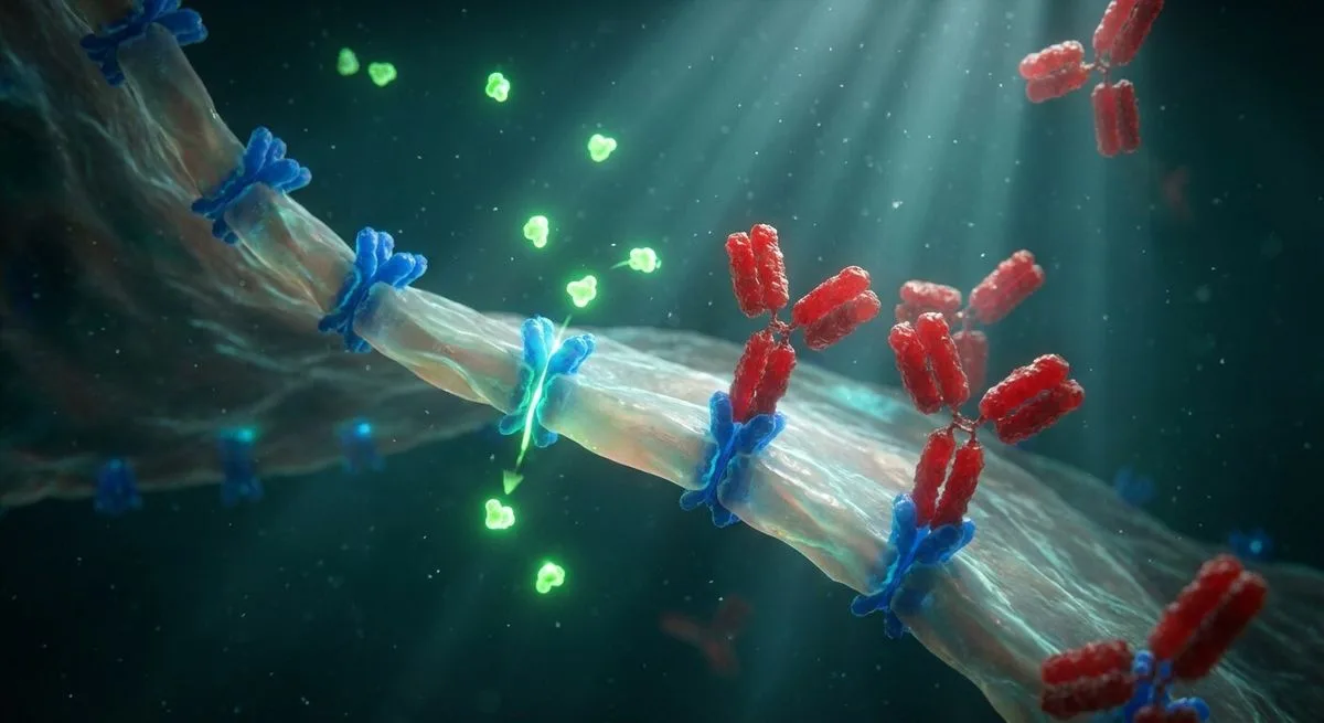
The brain and the bloodstream are separated by the blood-brain barrier. To cross it, folate needs a specific transporter — folate receptor alpha (FRα), located on the cells of the choroid plexus. If this receptor stops working, folate stays in the blood and the brain goes hungry. A standard blood test will never catch that.
FRα Antibodies: An Invisible Barrier
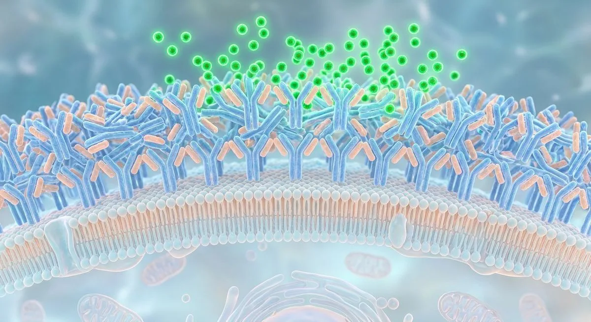
The most common reason FRα stops working is autoantibodies. The immune system produces antibodies that attack the body's own folate receptor.
Where do they come from? The FRα protein found in cow's milk shares about 90% of its amino-acid sequence with the human version. In genetically predisposed people, the immune system starts producing antibodies against the milk FRα — and those antibodies cross-react with the body's own receptor at the blood-brain barrier.
Two types of antibodies, two mechanisms of damage:
- Blocking antibodies — attach directly to the site where folate binds the receptor, preventing the vitamin from "docking"
- Binding antibodies — attach elsewhere but activate the complement system, which destroys the entire receptor complex
The result is the same either way: folate never reaches the brain.
The proof of this mechanism is simple and elegant: a dairy-free diet lowers FRα antibody titers within 3–6 months. Reintroduce milk, and the antibodies climb right back up.
There's another quirk: antibody titers fluctuate in cycles of roughly 5–7 weeks. That explains why a single negative test doesn't rule out the diagnosis — a repeat test a few weeks later can come back positive.
Age Windows: What to Look For, and When

The clinical picture of CFD depends on the age at which the antibodies start doing damage. Each age range has its own characteristic syndrome.
Before Birth
If the mother carries FRα antibodies, they disrupt folate transport across the placenta. Consequences include a higher risk of neural tube defects, autism, and developmental delay.
If the father carries the antibodies, they affect folate-dependent processes in sperm production, epigenetic programming, and DNA quality.
4–6 Months: Infantile CFD
The first signs are sleep disturbances, restlessness, and agitation that can't be explained by reflux or allergies. Over the following two years:
- Head circumference growth slows
- Psychomotor development is delayed, with hypotonia and ataxia
- Pyramidal signs appear in the lower limbs (without treatment, progressing to tetraspasticity)
- About a third of children develop dyskinesias and seizures
- Vision loss can begin from age 3, hearing loss from age 6
A minority go on to develop autism. On MRI, about half of untreated children show delayed myelination, demyelination, and atrophy of the brain and cerebellum.
Why this window matters so much: children who start folinic acid treatment before age 2 show the best outcomes — in some cases, full recovery.
1–2 Years: Spastic-Ataxic Syndrome
Movement and learning impairments appear when FRα antibodies develop at this age.
2–5 Years: ADHD
In some children with attention-deficit/hyperactivity disorder (ADHD), learning difficulties, and behavioral problems, FRα autoimmunity is found. Neurologically, this shows up as mild gait ataxia and a positive Romberg test.
Teens and Adults: The Psychiatric Spectrum
- Treatment-refractory schizophrenia: FRα antibodies found in 85% of patients (20 tested). The 5–7-week antibody cycle may explain the waxing-and-waning course of the illness.
- Treatment-resistant depression: FRα antibodies found in 56% (9 of 16 patients).
- Dystonia and parkinsonism: sporadic cases that don't respond to standard therapy.
Later in Life: Dementia
One case report describes dementia with myoclonus linked to CFD in an adult woman. No systematic studies exist yet.
Treatment Before Age 2 = A Chance at Full Recovery

Observational data on children with infantile CFD are unambiguous: the earlier treatment starts, the better the outcome.
Children who start folinic acid before age 2 can recover fully — development normalizes and neurological symptoms disappear. After age 6, the prognosis is significantly worse: lost time means lost windows of neuroplasticity.
The backbone of therapy is high-dose folinic acid (dl-5-formyltetrahydrofolate: 0.5–1 mg/kg/day, up to 2 mg/kg/day if needed, capped at 50 mg/day). Levo-folinic acid is used at half those doses.
One critical rule: start at half the target dose. A sudden rise in brain folate triggers a surge in dopamine and serotonin synthesis. Receptors that were suppressed during the long period of deficiency haven't had time to adapt, and the result can be pronounced agitation and aggression. It takes about six weeks for a new equilibrium to settle in. If agitation is severe, a low dose of risperidone is prescribed temporarily, for 2–3 months.
Alongside folinic acid, a dairy-free diet is introduced (replacing cow's milk with plant-based alternatives). It removes the source of the cross-reacting FRα protein and lowers antibody titers within 3–6 months. The diet can start right after diagnostic samples are collected, without waiting for results.
What Else Affects How Well Treatment Works
- Vitamin D: a deficiency reduces expression of RFC1, an alternative folate transporter. Correcting it is essential.
- MTHFR mutations: in the C677T homozygous or C677T/A1298C compound-heterozygous variant, part of the folinic acid dose is replaced with levo-methyl-THF, plus high-dose riboflavin (B2).
- Antioxidants and trace elements: deficiencies in vitamins C and E, coenzyme Q10, iron, copper, zinc, manganese, and selenium worsen oxidative stress, which further degrades folate and FRα. Comprehensive correction is needed.
Three Families, Five Healthy Children
This is the kind of data that could change how at-risk families approach planning a pregnancy.
In three families, the first child had autism. All the children tested positive for FRα antibodies. Genetic testing for known causes of autism (CNVs and associated genes) came back normal.
Researchers then tested the parents for FRα antibodies.
In all three families, the mothers tested positive. The plan: give both parents low-dose folinic acid (up to 3.75 mg/day of levo-folinic acid) starting three months before conception. Mothers with antibodies continued taking it throughout pregnancy.
The result:
- Family I: two healthy daughters and one healthy son
- Family II: one healthy daughter
- Family III: one healthy son
Five children, all healthy. Not a single case of autism.
Five children is not a randomized controlled trial. The authors of the review — Ramaekers and Quadros — are candid that this data is preliminary and needs confirmation in clinical studies. But they also note that preventing CFD and autism in offspring is a "real possibility, provided prospective parents are screened for FRα antibodies and associated genes and receive folinic acid treatment before conception."
Why the Prognosis Depends on the Parents' Antibodies
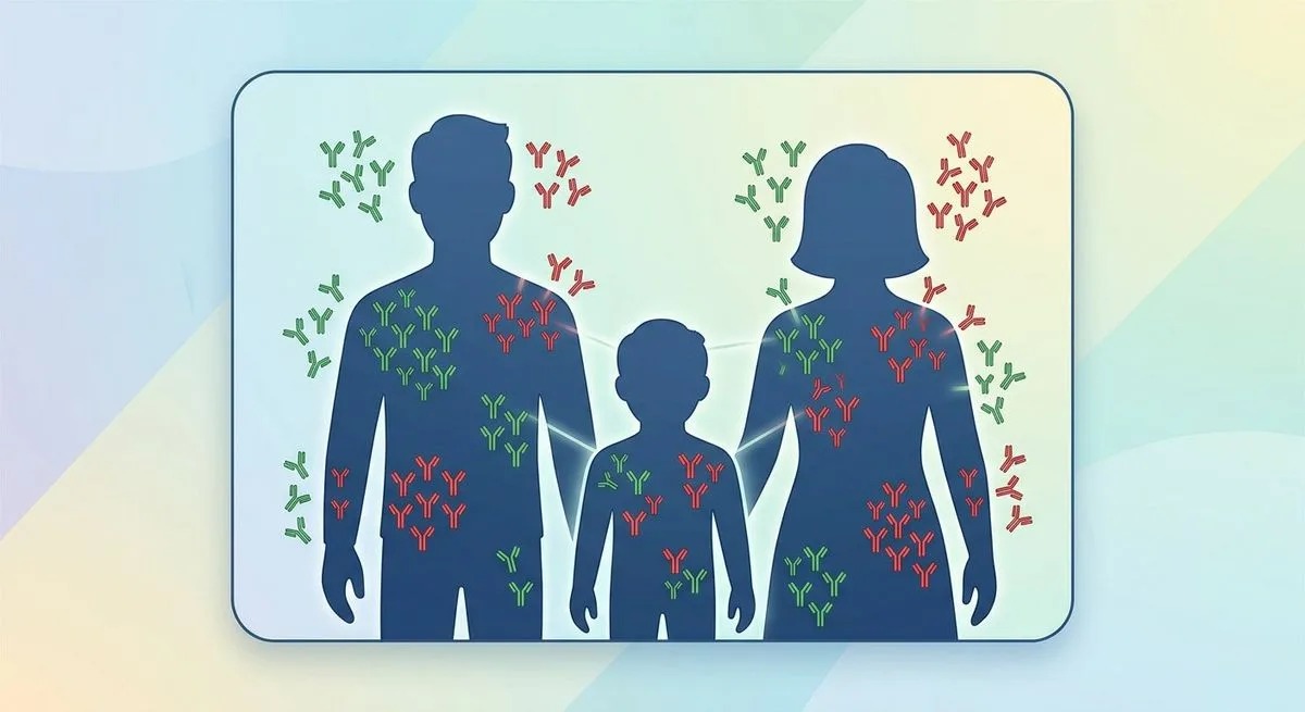
In autism, the prognosis for folinic acid treatment depends not only on when therapy starts, but also on the family's "antibody profile."
There are eight possible combinations of FRα antibody presence or absence across the child, mother, and father. The key scenarios:
-
Antibodies in the child only (both parents negative). The autism is acquired — antibodies developed after birth, likely triggered by cow's milk. Early folinic acid treatment plus a dairy-free diet gives the most favorable prognosis.
-
Antibodies in the child and one parent. Beyond the child's postnatal folate deficiency, there's exposure at the level of the gametes or during fetal development. Autism is partially reversible with treatment.
-
Antibodies in the child and both parents. This is the maximum exposure: folate-dependent spermatogenesis, folate-dependent oocyte maturation, fetal folate transport, and postnatal folate transport to the brain are all disrupted. The prognosis is poor even with treatment.
What Parents and Doctors Can Do Now
For Doctors: When to Suspect CFD
- A 4–6-month-old infant with unexplained restlessness, sleep disturbances, and agitation
- Developmental delay and slowing head-circumference growth despite normal standard labs
- Autism, especially with a normal genetic screen
- ADHD with mild ataxia
- Treatment-refractory schizophrenia, especially with a waxing-and-waning course
- Treatment-resistant depression
- Dystonia or parkinsonism that doesn't respond to standard therapy
The first step is a blood test for FRα autoantibodies (both blocking and binding types). Keep the antibody cycle in mind: if suspicion remains high despite a negative result, retest a few weeks later. Folate-containing supplements should be stopped 3 days before testing.
For Families With a Child on the Autism Spectrum

- Talk to a doctor about whether testing your child for FRα antibodies makes sense
- If the result is positive, both parents should be tested too — it affects the prognosis
- Seek genetic counseling before planning another pregnancy
The Future of Screening
The review's authors suggest discussing:
- FRα antibody screening in pregnant women and in cord blood
- Monitoring children for FRα antibodies at 6, 12, and 24 months
- Preventive folinic acid treatment for antibody-positive parents before conception
None of this is standard practice yet — but it points toward an approach that could reshape how we prevent neurodevelopmental disorders. Clinical studies to confirm or refute these proposals are still needed.